


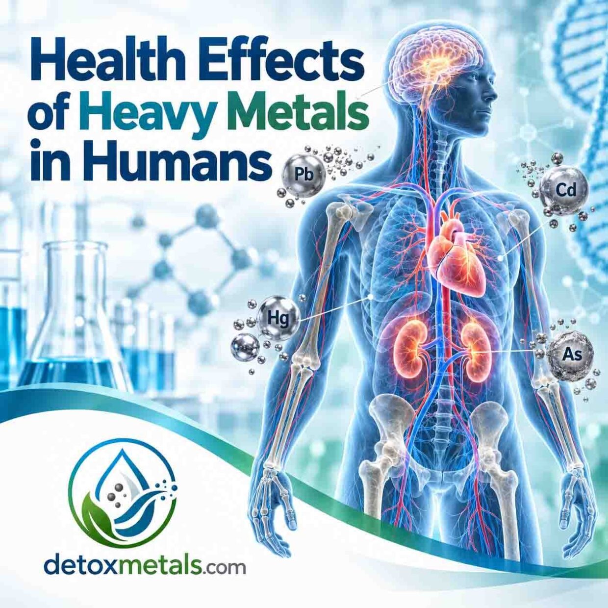
Health Effects of Heavy Metals
Health Effects of Heavy Metals in Humans
How lead, mercury, cadmium and arsenic affect the body, who is most vulnerable, how exposure is tested and what can be done to reduce risk
Learn how lead, mercury, cadmium and arsenic affect the brain, kidneys, heart, blood, bones and development, and how exposure is tested.
Heavy metals can enter the body through contaminated food or water, polluted air, tobacco smoke, consumer products, medicines, hobbies and the workplace. Their health effects depend on the metal and its chemical form, the dose, the route and duration of exposure, and the person’s age, nutritional status and health.
The most important first steps are to identify the source, stop or reduce exposure and use the right clinical test. A non-specific symptom on its own does not prove metal toxicity.
Suspected acute poisoning, breathing difficulty, confusion, seizures, severe weakness, persistent vomiting, severe abdominal pain or a known high-dose exposure requires urgent medical assessment or contact with a poison centre.
What Are Toxic Heavy Metals?
The term heavy metals is widely used in medicine and environmental health, although it is not a precise chemical category. It usually refers to dense metals and related elements that can cause toxicity at relatively low exposure.
Arsenic is technically a metalloid, but it is commonly discussed with toxic metals because its exposure pathways and health effects overlap.
Lead, mercury, cadmium and inorganic arsenic are among the substances of greatest public-health concern. They have no useful nutritional role in the human body. Exposure does not automatically mean illness: risk rises with the absorbed dose and with repeated or prolonged exposure.
Some forms are more hazardous than others. Methylmercury, for example, is the main form that accumulates in fish, while inorganic mercury salts and elemental mercury vapour behave differently in the body.
Organic arsenic compounds in most seafood are generally far less toxic than inorganic arsenic in contaminated water or food. [1-10]
How Heavy Metals Damage Cells
Toxic metals can damage cells through several connected mechanisms. Many bind to sulphur-containing groups in proteins, changing the shape and function of enzymes. They can displace essential minerals such as calcium, zinc, iron and selenium, disturb cell signalling and impair the production of haem, the iron-containing part of haemoglobin. [9-11]
They can also increase oxidative stress. This happens when reactive oxygen species are produced faster than antioxidant systems can control them. The resulting damage can affect cell membranes, proteins, mitochondria and DNA.
Mitochondria are especially important because they generate most of the cell’s usable energy. Metal-induced mitochondrial dysfunction may therefore contribute to fatigue, nerve injury and organ damage, although symptoms such as fatigue have many more common causes and are not diagnostic of metal toxicity. [9,10]
Some metals imitate normal ions and cross biological barriers. Lead can interfere with calcium-dependent processes; methylmercury can reach the brain and cross the placenta; cadmium accumulates mainly in the kidneys; and inorganic arsenic can alter cellular signalling, DNA repair and gene regulation. Chronic inflammation and impaired blood-vessel function can amplify the damage. [1-10]
Effects on the Brain and Nervous System
The developing brain is highly sensitive to toxic exposure. Lead can impair attention, learning, behaviour and intellectual development in children, even when poisoning is not obvious. Very high exposure can cause encephalopathy, seizures, coma and death. In adults, lead exposure has been associated with headache, mood or cognitive changes and peripheral nerve dysfunction. [1,5,11]
Methylmercury is a potent neurotoxicant. Prenatal exposure can affect the child’s cognitive, language, attention, memory and motor development. Adults with substantial mercury exposure may develop tremor, sensory disturbance, poor coordination, irritability, sleep disturbance or memory problems.
Chronic arsenic exposure can cause symmetrical peripheral neuropathy with numbness, tingling, pain or weakness. Cadmium is less classically neurotoxic, but high or chronic exposure may contribute indirectly through oxidative stress and kidney-related metabolic disturbance. [2,3,6-10]
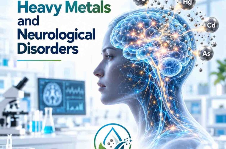
Read more: Heavy Metals and Neurological Disorders
Effects on the Kidneys
The kidneys filter the blood and concentrate many substances for excretion, which makes them vulnerable to toxic metals. Cadmium is a major renal toxicant. It accumulates in the proximal tubules, where long-term exposure can cause loss of small proteins, glucose, phosphate and other nutrients into the urine. Once substantial tubular damage has occurred, it may be irreversible. [4,7]
Lead exposure is associated with reduced kidney function and can contribute to chronic kidney disease, especially after prolonged or higher exposure. Inorganic mercury compounds can damage renal tubules and glomeruli, sometimes producing protein in the urine or acute kidney injury. Arsenic can also affect kidney function, particularly during significant acute exposure. [1-8]
Effects on the Cardiovascular System
Lead has one of the clearest cardiovascular links. Long-term exposure is associated with higher blood pressure and greater risk of cardiovascular disease. Proposed mechanisms include oxidative stress, reduced nitric-oxide availability, altered autonomic control, kidney damage and changes in the renin-angiotensin system. [1,12]
Chronic arsenic exposure in drinking water has been associated with cardiovascular disease and changes in blood-vessel function. Cadmium exposure has also been associated with coronary heart disease, stroke and peripheral arterial disease in epidemiological studies. These studies show population-level associations; an individual’s cardiovascular disease should not be attributed to a metal without an exposure history and appropriate testing. [3,13]
Effects on the Blood
Lead interferes with several enzymes needed to make haem. This can shorten red-cell survival and contribute to anaemia, particularly at higher exposures. A blood film may show basophilic stippling in significant, prolonged poisoning, but this finding is neither sensitive nor specific enough to diagnose lead exposure. [1,5,11]
Arsenic poisoning can affect bone marrow and may cause anaemia, low white-cell counts or low platelets. Cadmium may contribute to anaemia through kidney damage, impaired iron handling or direct toxic effects. A full blood count can reveal a problem, but it does not identify the responsible metal; metal-specific testing is needed. [3,7,16]
Effects on Bones
Lead is stored mainly in bone and teeth, where it can remain for years or decades. Blood lead can fall after external exposure stops, while bone stores persist. Periods of increased bone turnover, including pregnancy, breastfeeding, menopause or prolonged immobility, can release some stored lead back into the bloodstream. [1,5]
Cadmium can weaken bone both directly and through kidney damage that alters calcium, phosphate and vitamin D metabolism. Long-term exposure has been linked with osteomalacia, lower bone mineral density, osteoporosis and fracture risk. Adequate nutrition supports bone health, but supplements do not replace exposure control. [4,7,14]
Effects on Reproduction and Development
The embryo, fetus, infant and young child are especially sensitive because organs are developing rapidly and protective systems are immature. Lead crosses the placenta. Maternal exposure is associated with reduced fetal growth, preterm birth and harm to neurodevelopment. Lead exposure can also impair male and female reproductive function at sufficient doses. [1,5]
Methylmercury crosses the placenta and can affect the developing nervous system even when the mother has few symptoms. This is why pregnancy guidance usually focuses on choosing lower-mercury fish rather than avoiding fish entirely, since fish also provides protein and omega-3 fatty acids.
Arsenic exposure has been associated with adverse pregnancy outcomes, infant mortality and later developmental effects. Cadmium may affect placental function, fetal growth and reproductive health, although risk varies with dose and route. [2,3,6-8,15]
Lead – Main Health Effects
Lead can affect almost every organ system. Children absorb a greater proportion of ingested lead than adults and are more likely to ingest contaminated dust through normal hand-to-mouth behaviour. No safe blood lead concentration has been identified for children. [1]
- Brain and development: reduced attention, learning difficulties, behavioural effects and lower cognitive performance; severe exposure can cause encephalopathy, seizures or coma.
- Blood: inhibition of haem synthesis and anaemia at higher exposures.
- Kidneys and cardiovascular system: impaired renal function, raised blood pressure and increased cardiovascular risk with long-term exposure.
- Reproduction and pregnancy: reduced fertility, miscarriage risk, preterm birth, reduced fetal growth and developmental harm, depending on exposure.
- Bones: long-term storage with possible release during periods of increased bone turnover.
Common sources include deteriorating lead paint and contaminated household dust, renovation of older buildings, lead plumbing or fittings, battery manufacture and recycling, mining and smelting, firing ranges and ammunition, stained-glass work, some ceramics, cosmetics, spices and traditional remedies. [1,18]
Mercury – Main Health Effects
Mercury exists as elemental mercury, inorganic mercury compounds and organic forms such as methylmercury. The form and route matter. Methylmercury exposure occurs mainly through contaminated fish and shellfish. Elemental mercury vapour may be inhaled in some workplaces or after spills involving mercury-containing equipment. [2,6]
- Nervous system: tremor, altered sensation, poor coordination, memory or mood changes and, at high exposure, severe neurological injury.
- Development: prenatal methylmercury exposure can impair cognitive and motor development.
- Kidneys: inorganic mercury can cause proteinuria and renal injury.
- Lungs and digestive tract: substantial inhalation of mercury vapour can injure the lungs; inorganic salts can damage the gastrointestinal tract.
Fish remains a valuable food. Risk reduction means following local guidance, varying the species eaten and choosing lower-mercury options, especially during pregnancy and childhood. Dental amalgam contains elemental mercury, but removal solely for detoxification is not generally recommended in people with sound fillings; drilling can temporarily increase exposure and should be discussed with a qualified dental professional. [2,15]
Cadmium – Main Health Effects
Cadmium has a long biological half-life and accumulates mainly in the kidneys. Tobacco smoke is a major avoidable source. For non-smokers, diet is usually the main route, with exposure varying according to soil, food type and local conditions. Occupational exposure can occur in battery production, metal work, pigments, plastics and recycling. [4,7]
- Kidneys: proximal tubular damage, low-molecular-weight protein loss and declining renal function.
- Bones: loss of minerals, osteomalacia, osteoporosis and increased fracture risk.
- Lungs: inhaled cadmium fumes or dust can cause severe lung injury; long-term occupational inhalation raises cancer risk.
- Cardiovascular system: epidemiological studies associate cadmium burden with cardiovascular and peripheral arterial disease.
Cadmium and cadmium compounds are classified as carcinogenic to humans. The strongest prevention measures are avoiding tobacco smoke, controlling workplace exposure and following food-safety advice rather than trying to eliminate individual nutritious foods without evidence of contamination. [4,13,17]
Arsenic – Main Health Effects
Inorganic arsenic is the principal toxic form of concern. The most important global source is contaminated groundwater used for drinking, cooking and irrigation. Other sources can include mining and smelting, combustion, certain pesticides, pressure-treated wood and contaminated foods. Organic arsenic from seafood can raise total urinary arsenic without indicating toxic inorganic exposure. [3,8]
- Acute exposure: vomiting, abdominal pain, diarrhoea, dehydration, cardiovascular instability, encephalopathy and peripheral neuropathy.
- Skin: pigment changes and thickened areas on the palms and soles after chronic exposure.
- Nervous system: numbness, tingling, pain or weakness from peripheral neuropathy.
- Cardiometabolic effects: associations with cardiovascular disease and diabetes in chronically exposed populations.
- Cancer: chronic inorganic arsenic exposure causes cancers of the skin, lung and bladder and is linked with other cancers.
Because chronic arsenic toxicity can develop slowly, the exposure history is essential. Testing drinking water is often more useful for prevention than repeatedly testing people after the source has already been identified. [3,8,17]
Who Is Most Vulnerable?
Vulnerability is determined by both exposure and biology. The groups that require particular care include:
- Fetuses, infants and young children, because the brain and other organs are developing.
- Pregnant or breastfeeding people, because some metals cross the placenta or enter breast milk and bone lead may be remobilised.
- Workers in mining, smelting, welding, battery manufacture or recycling, construction, renovation, firing ranges, electronics recycling and other metal-related industries.
- People living near mines, smelters, waste sites or industrial emissions, or using private wells in areas with naturally high arsenic.
- Smokers and people regularly exposed to second-hand smoke, because tobacco is an important cadmium source.
- People with kidney disease, iron deficiency, calcium deficiency or poor nutrition, which may increase absorption or reduce resilience.
- People using imported cosmetics, spices, pottery, traditional remedies or supplements from sources with inadequate quality control.
When Should Heavy Metal Exposure Be Investigated?
Investigation is most useful when symptoms are paired with a credible exposure route. Consider a clinician-led exposure assessment when there is known workplace or environmental contact; use of contaminated water or a recalled product; renovation of an older property; frequent firing-range use; home smelting, stained glass or battery work; mercury spills; pica in a child; or unexplained similar illness in household members or co-workers.
Possible clinical clues include unexplained anaemia, kidney tubular abnormalities, peripheral neuropathy, tremor, cognitive or behavioural change in a child, persistent gastrointestinal symptoms after a suspected exposure, characteristic arsenic-related skin changes or an unusual cluster of symptoms after a specific event.
These findings are not specific. Fatigue, brain fog, headache and digestive symptoms are common in many conditions, so assuming heavy metals are the cause can delay the correct diagnosis.
Testing
Testing should be chosen for the metal, the likely route and the timing of exposure. A clinician should begin with a detailed history covering work, hobbies, home renovation, water source, diet, smoking, medicines, supplements, imported products and where household members spend time. Environmental testing may be needed to find the source. [16,18]
- Lead: a venous blood lead level is the standard test for recent or ongoing exposure. Capillary results that are elevated usually need confirmation with a venous sample. Blood lead may underestimate a remote body burden because much lead is stored in bone. [18]
- Mercury: whole blood is often used for recent methylmercury exposure; urine is generally more useful for elemental or inorganic mercury exposure. Interpretation depends on mercury species, fish intake and timing. [2,6]
- Cadmium: blood cadmium reflects recent exposure, while urinary cadmium, usually adjusted for creatinine, is more useful for long-term body burden. Kidney markers may be added when chronic exposure is suspected. [7,19]
- Arsenic: a 24-hour urinary speciated arsenic test is the most informative test for recent exposure because it separates inorganic arsenic and its metabolites from less toxic seafood-derived forms. Seafood can raise total urine arsenic for about 48 hours. [16]
Hair and nail analysis can be affected by external contamination, cosmetic treatment, laboratory methods and the absence of accepted clinical reference ranges for many metals. These samples may have a role in specialised forensic or research settings, but they should not be used alone to diagnose toxicity or decide treatment.
Similarly, post-chelator or provoked urine testing cannot be compared with ordinary, non-provoked reference ranges and is not recommended as a routine diagnostic method by the American College of Medical Toxicology. [16,18,20]
A result above a reference range confirms exposure, not necessarily that the metal caused every symptom. Interpretation should combine the laboratory result with the exposure history, examination, kidney and liver function, full blood count and other tests selected for the patient.
Reducing Exposure
The most effective intervention is to stop exposure. Supplements cannot compensate for contaminated water, unsafe work practices or continuing contact with a toxic source.
- Test and treat the source: use an accredited laboratory for private-well water or suspect products, and follow local public-health advice.
- Use workplace controls: ventilation, wet methods, suitable respiratory protection, protective clothing, handwashing and changing before returning home. Avoid bringing contaminated dust home on shoes or work clothes.
- Manage older buildings safely: use trained contractors for lead paint or dust and avoid dry sanding or uncontrolled demolition.
- Do not smoke, and reduce exposure to second-hand smoke.
- Choose fish according to current local pregnancy and food-safety guidance; vary species and favour lower-mercury choices.
- Buy medicines, cosmetics, spices, pottery and supplements from traceable suppliers with credible quality control.
- Maintain a balanced diet with adequate iron, calcium, zinc, selenium and protein. Deficiency can increase the absorption or effects of some metals, but high-dose supplementation can also be harmful.
Medical chelation is reserved for selected, confirmed poisonings and must be supervised by a clinician experienced in toxicology. Chelators can cause serious adverse effects and can remove essential minerals. The correct agent, dose and timing depend on the metal, the exposure level and the patient’s condition. [16,18,20]
HMD® Triple-System Protocol
The HMD® Tri-Active Protocol is a complementary supplement programme developed around three stages: HMD® as the core formulation and intracellular eliminator, HMD® Chlorella for the gut-binding stage, and HMD LAVAGE® to support normal detox organs and elimination pathways. The programme describes these stages as mobilisation, binding and elimination support.
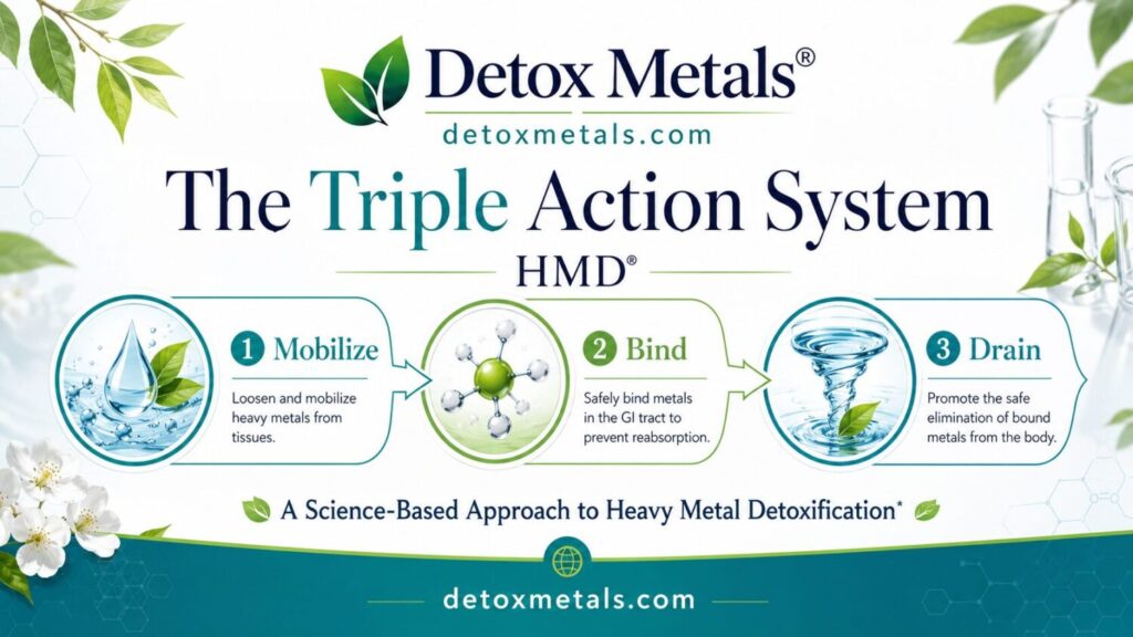
The rationale is practical: a detox programme should consider both the movement of compounds and their exit through the gastrointestinal and urinary systems. However, these proposed actions should not be treated as proof that the products remove a diagnosed toxic body burden, prevent organ damage or treat poisoning.
The developer-authored 2018 paper describes controlled testing of several natural substances in metal-exposed workers and subsequent assessments of the HMD formulation. It is an initial body of evidence, and larger independent trials with clinically meaningful outcomes are still needed. [21]
For responsible use, begin with exposure control and appropriate medical testing. HMD® should not replace urgent care, poison-centre advice or clinician-directed chelation. Children, pregnant or breastfeeding people, and anyone with kidney or liver disease, significant medical conditions or regular medication should seek professional advice before using a detox supplement.
Stop the product and obtain advice if new or worsening symptoms occur. Product use should follow the current label and practitioner guidance.
A structured programme is most useful when it sits within a wider plan: remove the source, support adequate nutrition and hydration, maintain regular bowel function, monitor relevant clinical markers when indicated and reassess whether the original exposure has actually fallen. The goal is safe, measurable risk reduction rather than attributing every non-specific symptom to heavy metals.
Scientific References
- World Health Organization. Lead poisoning and health. Updated 10 June 2026. https://www.who.int/news-room/fact-sheets/detail/lead-poisoning-and-health
- World Health Organization. Mercury and health. Updated 24 October 2024. https://www.who.int/news-room/fact-sheets/detail/mercury-and-health
- World Health Organization. Arsenic. Updated 7 December 2022. https://www.who.int/news-room/fact-sheets/detail/arsenic
- World Health Organization. Cadmium. Chemical Safety and Health. https://www.who.int/teams/environment-climate-change-and-health/chemical-safety-and-health/health-impacts/chemicals/cadmium
- Agency for Toxic Substances and Disease Registry. Toxicological Profile for Lead. Atlanta: US Department of Health and Human Services; 2020. https://www.atsdr.cdc.gov/toxprofiles/tp13.pdf
- Agency for Toxic Substances and Disease Registry. Toxicological Profile for Mercury. Atlanta: US Department of Health and Human Services; 2024. https://www.atsdr.cdc.gov/toxprofiles/tp46.pdf
- Agency for Toxic Substances and Disease Registry. Toxicological Profile for Cadmium. Atlanta: US Department of Health and Human Services; 2012. https://www.atsdr.cdc.gov/toxprofiles/tp5.pdf
- Agency for Toxic Substances and Disease Registry. Toxicological Profile for Arsenic. Atlanta: US Department of Health and Human Services; 2007. https://www.atsdr.cdc.gov/toxprofiles/tp2.pdf
- Tchounwou PB, Yedjou CG, Patlolla AK, Sutton DJ. Heavy metal toxicity and the environment. EXS. 2012;101:133-164. https://doi.org/10.1007/978-3-7643-8340-4_6
- Jaishankar M, Tseten T, Anbalagan N, Mathew BB, Beeregowda KN. Toxicity, mechanism and health effects of some heavy metals. Interdiscip Toxicol. 2014;7(2):60-72. https://doi.org/10.2478/intox-2014-0009
- Flora G, Gupta D, Tiwari A. Toxicity of lead: a review with recent updates. Interdiscip Toxicol. 2012;5(2):47-58. https://doi.org/10.2478/v10102-012-0009-2
- Navas-Acien A, Guallar E, Silbergeld EK, Rothenberg SJ. Lead exposure and cardiovascular disease: a systematic review. Environ Health Perspect. 2007;115(3):472-482. https://doi.org/10.1289/ehp.9785
- Tellez-Plaza M, Jones MR, Dominguez-Lucas A, Guallar E, Navas-Acien A. Cadmium exposure and clinical cardiovascular disease: a systematic review. Curr Atheroscler Rep. 2013;15(10):356. https://doi.org/10.1007/s11883-013-0356-2
- Engström A, Michaëlsson K, Vahter M, Julin B, Wolk A, Åkesson A. Associations between dietary cadmium exposure and bone mineral density and risk of osteoporosis and fractures among women. Bone. 2012;50(6):1372-1378. https://doi.org/10.1016/j.bone.2012.03.018
- National Research Council. Toxicological Effects of Methylmercury. Washington, DC: National Academies Press; 2000. https://doi.org/10.17226/9899
- Agency for Toxic Substances and Disease Registry. Clinician Brief Arsenic. https://www.atsdr.cdc.gov/environmental-medicine/hcp/clinician-briefs/arsenic.html
- International Agency for Research on Cancer. Arsenic, Metals, Fibres and Dusts. IARC Monographs, Volume 100C. Lyon: IARC; 2012. https://publications.iarc.fr/120
- Agency for Toxic Substances and Disease Registry. Lead Toxicity Clinical Assessment Diagnostic Tests and Imaging. https://archive.cdc.gov/www_atsdr_cdc_gov/csem/leadtoxicity/diagnostic_testing.html
- Agency for Toxic Substances and Disease Registry. Cadmium Toxicity Clinical Assessment Laboratory Tests. https://archive.cdc.gov/www_atsdr_cdc_gov/csem/cadmium/Laboratory-Evaluation.html
- American College of Medical Toxicology. ACMT recommends against use of post-chelator challenge urinary metal testing. J Med Toxicol. 2017;13(4):352-354. https://doi.org/10.1007/s13181-017-0624-6
- Georgiou G. Scientific research on natural heavy metal chelators: testing what works. Int J Complement Alt Med. 2018;11(5):262-267. https://doi.org/10.15406/ijcam.2018.11.00409
This article is for education and does not replace individual medical care